Finger osteoarthritis
Finger osteoarthritis causes pain, deformities, and stiffness. While they don't offer a complete cure, there are medical and surgical treatments—often overlooked—that improve or stabilize the functional and aesthetic consequences of finger osteoarthritis.
Contents:
What is finger osteoarthritis?
Finger osteoarthritis is a common condition, particularly affecting women. It causes pain, deformities, and stiffness with often significant functional and aesthetic consequences.
This page does not cover osteoarthritis affecting the base of the thumb (or thumb osteoarthritis), which affects the trapeziometacarpal joint and is discussed in a separate information page.
The consequences of finger osteoarthritis for patients, as well as the available treatment options, are often underestimated by the medical community, which can further increase the anxiety of those affected.
Finger osteoarthritis develops progressively, with an earlier or later onset depending on the patient. It may affect one or several joints, on a single finger or on multiple fingers of both hands. There is no strict rule.
Causes of finger osteoarthritis
The origin of osteoarthritis remains poorly understood and is multifactorial, although a genetic cause is highly probable. In some families, finger osteoarthritis appears to be “passed down” from mother to daughter.
Other factors may also play a role, such as trauma or fracture sequelae. Inflammatory diseases such as rheumatoid arthritis may also contribute.
Certain inflammatory diseases such as rheumatoid arthritis or gout may also lead to osteoarthritis.
Mechanism of osteoarthritis
Osteoarthritis is the destruction of the cartilage covering the ends of the finger bones at the level of the joint.
Cartilage allows each bony surface to glide smoothly against the other during movement while distributing mechanical stress evenly.

The joint is also composed of ligaments connecting the two phalanges together and is surrounded by a capsule lined with a membrane that produces synovial fluid inside the joint. All of these structures are altered in osteoarthritis.
A distinction is made between primary osteoarthritis, related to aging, and post-traumatic osteoarthritis, resulting from mechanical joint injury after one or more repeated impacts. This subsequently causes progressive wear of the articular cartilage.
In the finger, there are 3 joints, named from the wrist toward the fingertip:
- metacarpophalangeal joint,
- proximal interphalangeal joint,
- distal interphalangeal joint.
The last two joints are the most commonly affected by finger osteoarthritis. Treatment differs depending on which joints are involved.
Diagnosis of finger osteoarthritis
The progression of finger osteoarthritis is generally gradual. One or several finger joints begin to swell and become painful. Initially, pain is not always constant and may occur in inflammatory flare-ups.
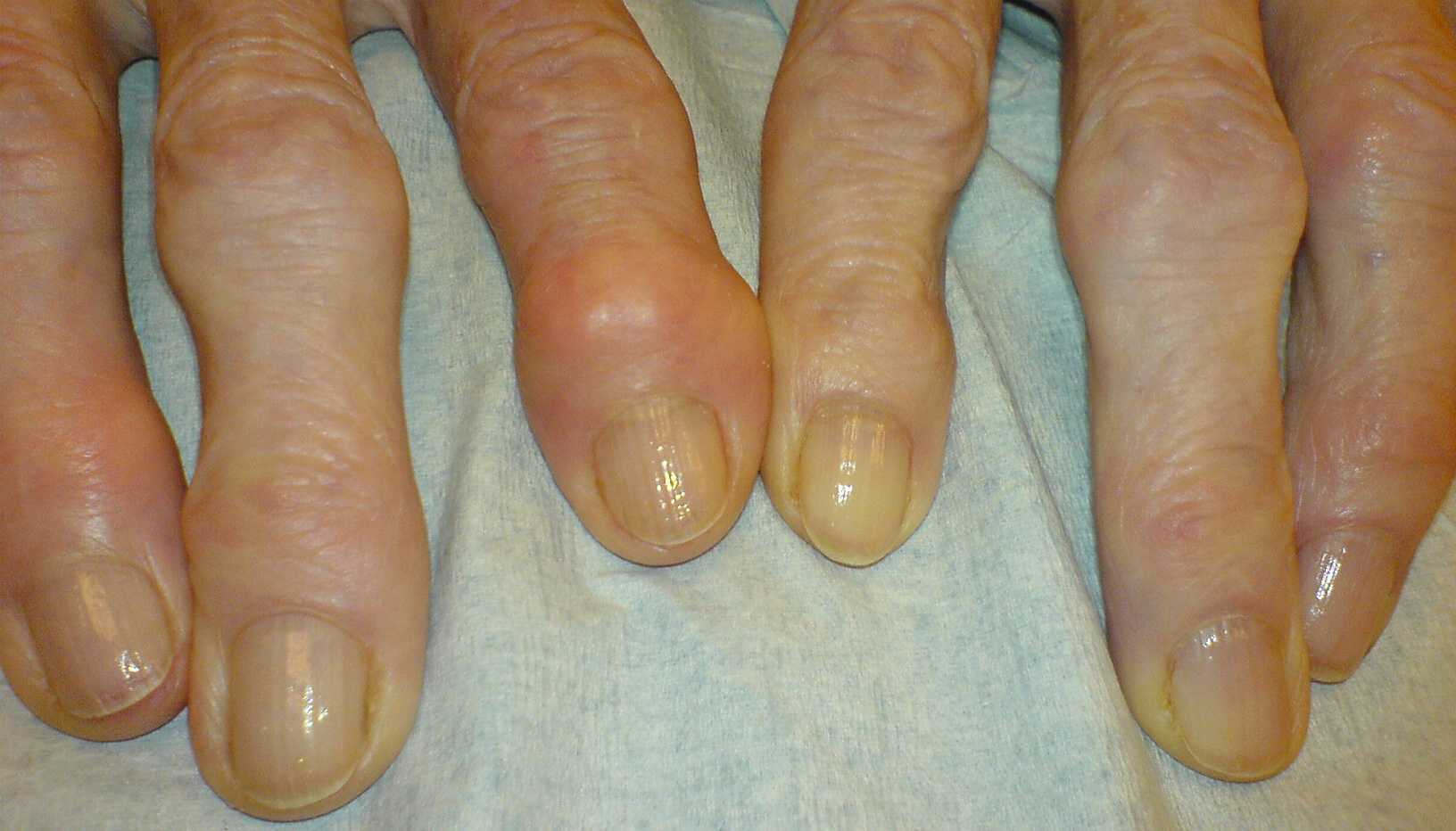
A deformity gradually appears. “Bumps” are often visible on the top and side of the joint, especially at the distal interphalangeal joint (the joint near the fingernail). These bumps are called Heberden’s nodes.
The proximal interphalangeal joint may also become deformed. In this case, the bumps are referred to as Bouchard’s nodes.
Stiffness also progressively develops, sometimes associated with angular deformity. The finger may begin to “deviate sideways.”
Other conditions may be associated with finger osteoarthritis, including the development of a mucous cyst, carpal tunnel syndrome, or tenosynovitis.
An X-ray is the standard examination and allows diagnosis and assessment of lesion severity.
A CT scan or MRI may sometimes also be requested.

Treatments for finger osteoarthritis
Treatment may be medical or surgical, depending on the stage of osteoarthritis, the functional, painful, and aesthetic impact, and the patient’s wishes.
No treatment is mandatory for a functional condition such as osteoarthritis. No treatment can provide a complete cure. However, although still not fully understood, several options exist to improve or stabilize the effects of osteoarthritis.
Medical treatment
- Painkillers, anti-inflammatory medications, and immobilization with a splint may be useful during painful flare-ups.
- Certain medications prescribed by rheumatologists may help slow the progression of osteoarthritis.
- Corticosteroid injections may also be prescribed during painful episodes.
- PRP injections, meaning growth factors taken from the patient’s own blood, are increasingly proposed depending on the stage of the disease, although consistent results cannot be guaranteed.
NOTE: As Dr. Roure is an orthopedic surgeon, he will only be able to manage your care if there is a surgical indication. In cases of early-stage osteoarthritis, it is recommended to consult a rheumatologist first for medical management.
Surgical treatment: when should it be considered?
Surgery may be considered when medical treatments have failed. It may also be proposed immediately in advanced and disabling forms of osteoarthritis.
Pain, stiffness, deformity, and aesthetic concerns are the main reasons to consider surgery.
Several surgical procedures may be proposed. They do not truly cure osteoarthritis or restore a completely normal finger, but they can significantly improve function, pain, and the appearance of the hand.
Depending on the stage of the disease and the expected outcome, 3 types of surgery may be considered to treat finger osteoarthritis surgically:
Joint resurfacing and debridement
Indication
Surgical resurfacing with joint debridement is proposed in the early stages of finger osteoarthritis. In these cases, the joint remains mobile and well aligned (without deviation). Osteoarthritis causes painful and unsightly joint deformity. Most often, the deformity is associated with bumps on the top of the joint (Heberden’s nodes).
Principle of the procedure
The operation consists of removing the bony overgrowths through two short incisions. A joint debridement and skin tightening are then performed. A PRP (growth factor) joint injection is generally carried out during the same procedure.
Results of resurfacing and debridement
The result can only be assessed after several months. The joint may remain swollen during the first weeks, followed by significant improvement in appearance and pain.
However, osteoarthritis cannot be considered cured. This is not possible with this technique. Finger osteoarthritis may continue to progress more or less rapidly depending on the patient.
In some advanced forms affecting the distal interphalangeal joint (fingertip joint), slight flexion of the finger may persist due to relaxation of the extensor tendon.
The main risk of the procedure is insufficient improvement compared to the patient’s expectations.
Summary
- Anesthesia: Regional
- Hospital stay: Outpatient (no overnight stay)
- Immobilization: Splint and dressing for about 2 weeks
- Rehabilitation: Self-rehabilitation by the patient; the finger can be used for daily activities from 2 weeks after surgery
Arthrodesis
Indications for arthrodesis in finger osteoarthritis
Arthrodesis is a procedure that consists of fusing the joint in a straight position.
This operation is generally proposed in the most advanced cases of finger osteoarthritis, where the joint is already very stiff. In the vast majority of cases, the surgery involves only the distal interphalangeal joint (the joint at the fingertip).
By fusing the two finger bones together, this technique allows for pain relief and improvement of the finger’s appearance. The procedure provides a straightened finger with reduced deformities and bony bumps.
This improvement comes at the cost of loss of mobility in the last phalanx.
In practice, finger function is often significantly improved with better grip. At this stage, strength and absence of pain are preferable to the residual mobility of the distal phalanx.
Procedure
A short incision is made on the back of the joint. The joint is fused after removal of the cartilage destroyed by osteoarthritis and elimination of the bony overgrowths.
To fuse the joint, pins are generally used until the two finger bones unite. Fusion usually occurs 6 to 8 weeks after surgery, with the use of a protective splint. The pins are then removed.
Summary
- Anesthesia: Regional
- Hospital stay: Outpatient (no overnight stay)
- Immobilization: Splint and dressing for about 6 to 8 weeks
- Rehabilitation: Self-rehabilitation by the patient; the finger can be used for daily activities after splint removal, while the other fingers can be used immediately
Joint prosthesis
Indications for joint replacement
Joint prosthesis surgery is limited to the proximal interphalangeal and metacarpophalangeal joints. Joint replacement is preferred over arthrodesis at these levels because fusion would cause significant functional impairment.
This surgical procedure for finger osteoarthritis is generally proposed in advanced osteoarthritis with pain and loss of motion.
Procedure
The operation consists of replacing the destroyed joint with a prosthesis. Depending on the indication, the prosthesis may be made of metal, silicone, pyrocarbon, or another synthetic material.
Placement of the prosthesis must be performed with precision. The earlier the surgery is performed, the better the expected outcome.
The goal of this procedure is to achieve a functional finger, with little or no pain, in a durable way. However, it should be understood that the range of motion obtained often remains below normal.
Summary
- Anesthesia: Regional
- Hospital stay: Outpatient (no overnight stay)
- Immobilization: Splint and dressing for about 2 weeks
- Rehabilitation: Self-rehabilitation by the patient and gentle physiotherapy starting 2 to 3 weeks after surgery. The finger can then gradually be used for daily activities, while the other fingers can be used immediately.
Possible complications
Complications following surgery for finger osteoarthritis are rare. However, recovery is often long. To achieve the best possible results, the patient must remain motivated for the surgery, immobilization, and rehabilitation.
Pain
Sometimes, residual pain during use may persist for several months after surgery. This pain is rarely present at rest.
Risks
Although rare and unpredictable, surgical procedures may involve risks. These risks must be weighed against the benefits compared with no treatment.
- Complex regional pain syndrome (CRPS) remains possible. This may result in pain and stiffness of the hand and wrist extending to the shoulder. The progression of this syndrome is often long and difficult and may leave permanent mobility limitations.
- Septic arthritis is rare but always possible. It may require a second surgical procedure or appropriate medical treatment.
- Depending on the skin type, inflammatory scarring may occur.
- In cases of arthrodesis, the two phalanges may sometimes fail to fuse completely, leading to persistent mobility at the joint. This is not problematic unless pain persists, in which case another bone graft procedure may be proposed.
- In cases of joint replacement, radiological signs of bone resorption are common and usually have no consequences. Prosthetic components may occasionally break because of their small size, requiring revision surgery.